Electrical Impedance Tomography
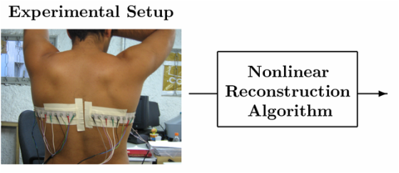
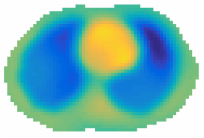
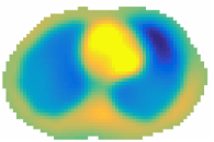
My main area of research involves the use of nonlinear inversion methods for an imaging modality called Electrical Impedance Tomography, or EIT for short. EIT is an imaging modality in which a conductive body is probed by harmless electrical currents applied through electrodes placed on its boundary. The currents penetrate the body to varying depths and the resulting boundary voltages are measured at the electrodes. From these surface measurements (voltages and current densities corresponding to knowledge of a Dirichlet-to- Neumann map), we aim to reconstruct an image of the internal conductivity and/or permittivity of the object. The schematic below demonstrates the setup; the reconstructions are computed for simulated EIT data using our nonlinear D-bar algorithm. The reconstruction task is a highly ill-posed nonlinear inverse problem and requires the use of mathematical techniques from functional and complex analysis, inverse scattering and PDE theory, and numerical analysis.
The conductivity and permittivity of biological tissues such as heart, lung, blood, and fat are different allowing a medical doctor to then use the EIT images as diagnostic tools. Medical applications include: monitoring heart and lung function in ICU patients, detection and classification of breast tumor, and brain imaging. Applications are not limited to medical fields alone and include: non-destructive evaluation, detection of groundwater contamination, oil exploration and landmine detection.
Recently, I have been exploring how to directly embed spatial a priori information into a nonlinear D-bar method. Including approximate organ boundaries known with high confidence (e.g. heart and lung positions extracted from a CT scan or atlas matching) can greatly sharpen EIT images.
|
|
The CGO sinogram
The traces of the CGO solutions obtained in the first step of D-bar approaches for the EIT problem contain more transparent information about the internal conductivity distribution than the voltage measurements or D-N map (see the illustrative video below). Furthermore, even partial data CGO traces can be recovered for low scattering frequencies. At the Centre of Excellence in Inverse Problems Research I have been working on a novel approach with Samuli Siltanen called a CGO sinogram, an analog of the sinogram used in X-ray tomography. The CGO sinogram is comprised of a matrix of the CGO boundary traces ranging over a circle |k| of fixed radius within the region of proven stability from regularization theory.
Phantom |
Dirichlet-to-Neumann Map |
CGO sinogram |
